
Designed for Pathology Minds Like Yours
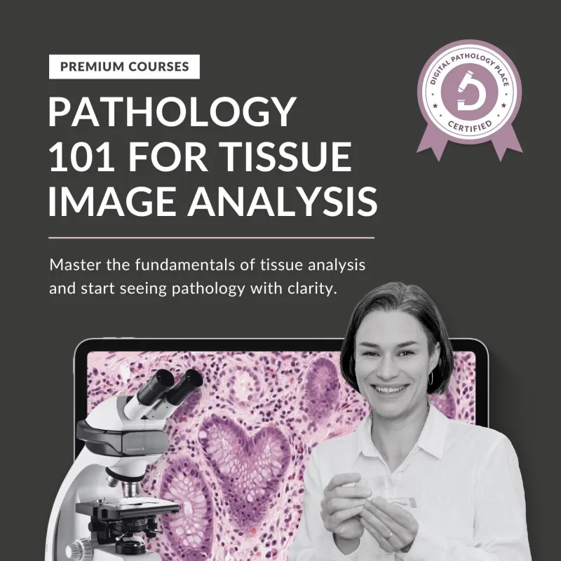


Pathology 101 For Tissue Image Analysis
$597.00
Quantity


© 2025 Digital Pathology Place - Aleksandra Zuraw, DMV, Ph.D., Dipl., ACVP
PO. Box 71, Fairfield, PA 17320, USA
Pathology 101 For Tissue Image Analysis
$597.00
Quantity
© 2025 Digital Pathology Place - Aleksandra Zuraw, DMV, Ph.D., Dipl., ACVP
PO. Box 71, Fairfield, PA 17320, USA